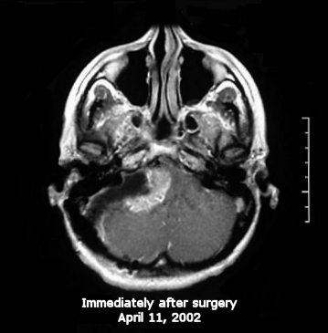
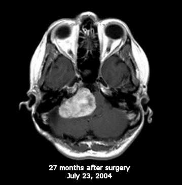
Follow- up image after surgery after tumor consolidation.
Just prior to radiation treatment.
This is just prior to the shunt surgery, 7
months after radiation.
Gray areas in the tumor show where the blood circulation is impaired because of tumor cell
death and consequently not taking up the dye that highlights the tumor.
It is uncertain whether the significant increase in size is due to further tumor growth,
or to edema* due to radiation insult/injury. If it is edema, it should subside on it's own
after a period of time - although that may be a year or more.
* (edema: swelling caused by an abnormal accumulation of fluid in body tissues)

Note the swelling of the tissues in her cheeks due to water retention from the Decadron steroid.
Dark areas in the brain adjacent to the tumor indicate
swelling and excess fluid from radiation damage to the
brain.
Small animated gif of comparison images 1MB file
Large animated gif of comparison images 6MB file