Note her right-deviated septum in her nose, the legacy of a teenage car wreck.
Val's "After" MRI images
(and some others)
| Here are Val's 2nd day post-surgery MRI images, put up next to the "before" images for comparison. I tried to select the ones that were most similar in cross section location to the first ones. Keep in mind they were done on a different day on a different machine and at slightly different planes through the head. Once again, all images have been mirrored so it is more intuitively obvious that the tumor is on Val's left side. |
|
|
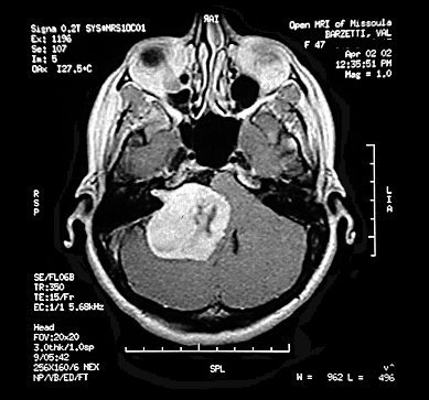
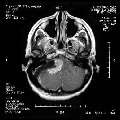
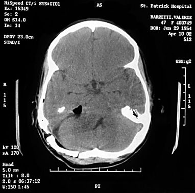
| Before - axial view, showing tumor in largest dimensions. The features within the tumor turned out to be an arterial branches critical to the brainstem. | After - axial view. This slice is tilted
slightly higher than the one to the left, and goes through the sinus cavities above and
behind the eyes. Note the brighter squiggly lines in what is left of the tumor. Those are
some of the critical arteries that caused the surgeons to leave so much of the tumor. The
dark gray area extending down and to the left of the remaining tumor is the path in from
the lower skull, and is filled with Cerebro-Spinal Fluid mixed with blood. Note her right-deviated septum in her nose, the legacy of a teenage car wreck. |
|
|
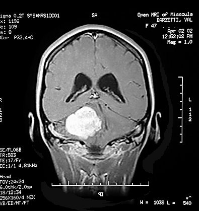
| Before - coronal view | After - coronal view This image is
sized so the skull is shown the same size as the "before" image to the left.
|
| Here is a CT image (better for bone detail) that shows the bone section that was removed and replaced. The dark area is an air pocket that will be absorbed into the blood and CSF. As an interesting side note, this bone flap is held in place by several small titanium tabs which are fastened to the bone with nine tiny titanium screws. Val can challenge the Pentagon procurement in this area, because she has an investment of almost a thousand dollars for those nine tiny screws. | |
 |
|
|
 |
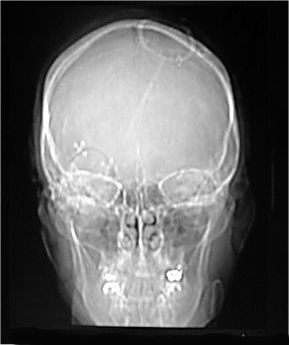
| This is a simple x-ray of Val's skull, part of the CT index series.
Besides being a sort of haunting image of a face/skull, is shows the CSF
shunt sewn into her scalp and penetrating her brain on the right side (avoiding the
speech center in the left upper brain) and crossing down through the centerline to the
left ventricle.
|
This is a CT index image, sagittal view, which shows the location and numbers of the axial views. It also shows the CSF shunt from the side - the faint line from the top of her skull in line with and pointed toward her spine. |
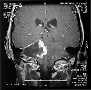
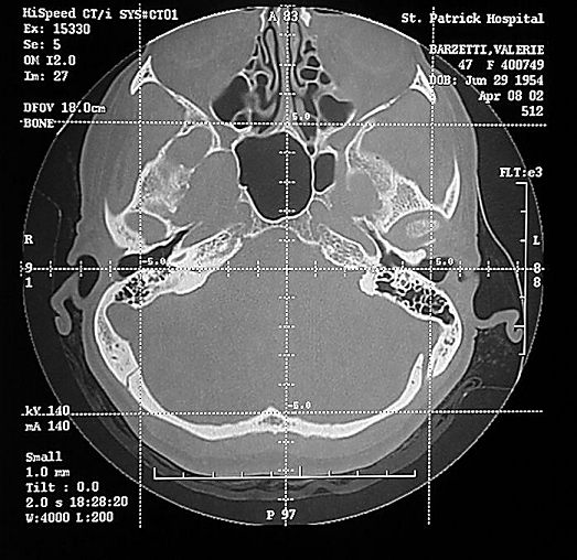
| There are many other fascinating and beautiful (to me, anyway) images in the several series of CT and MRI images. This last one is part of a pre-op study series Dr Von Doersten did to learn of Val's bone structure before he started cutting and drilling. It goes through a section of her mastoid (bone filled with air pockets behind the ear) and displays many asymmetries in her skull, and the delicacy of the structures in the upper nose and sinuses. | |
 |
|