Despite the larger size of the black background, the size of the skull image is precisely the same.
It was measured at 3.4 x 2.3 x 2.0 centimeters for a volume of 8.2 cubic centimeters
Val's 19 month MRI images
| For comparison, the 8 month and 19 month images are placed side by side. |
|
|
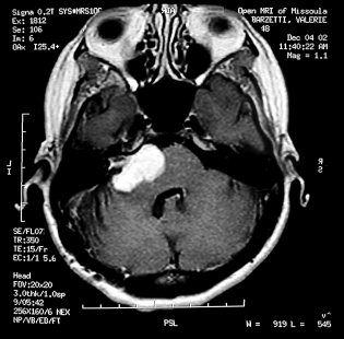
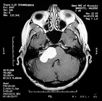
| 8 month axial image. | 19 month axial image at the same image slice position. Despite the larger size of the black background, the size of the skull image is precisely the same.
|
|
|
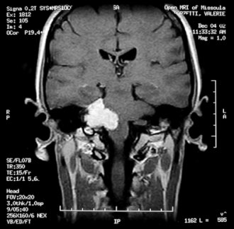
| 8 month coronal image. It was measured at 3.4 x 2.3 x 2.0 centimeters for a volume of 8.2 cubic centimeters |
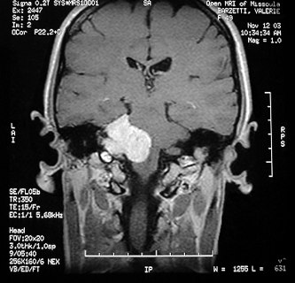
19 month coronal image. Dr. VonDoersten measured it at 3.8 x
2.4 x 2.9 centimeters for a volume of 13.2 centimeters, a 63% increase.
|
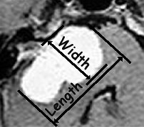
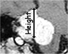
| The doctors measure the tumor in specific places and directions. From the literature and from watching the doctors, this is my understanding of those locations. | |
|
|
| Axial image width and length measure directions | Coronal image height measure location |